Sunday, October 18, 2015

Unit 3 Reflection

In Unit 3, we studied the different types and parts of cells, and their functions, and we focused specifically on cellular respiration and photosynthesis.

The different types of cells are eukaryotes and prokaryotes. Prokaryotes are cells that don't have a nucleus or most organelles. Some examples of prokaryotes include bacteria. Eukaryotes are cells with most organelles, including a nucleus. It is theorized that mitochondria and chloroplasts are prokaryotic cells that continued to live after being eaten by a eukaryotic cell.

Membranes of cells are made of 2 layers of lipids. Different membranes include in nuclear membrane, which holds the DNA in the nucleus. The lysosome digests old organelles that don't work any more, or old cells that don't work. There are 2 types of Endoplasmic Reticulum (ER), rough ER, which has ribisomes on the surface, and it helps finish making proteins. Smooth ER has no ribisomes on the surface, and detoxifies drugs. Vesicles take things out of cells. the Golgi Apparatus packages finished proteins, lipids, and hormones. Chloroplasts go through the process of photosynthesis to make glucose, and mitochondria break down glucose to create ATP for energy. And the cell membrane holds everything in the cell, and selects what enters and leaves. Other organelles are the nucleolus, which is the center of the nucleus, which starts ribisome production. In the nucleus, DNA is stored. Vacuoles store things sch as water, salts, proteins, and carbs, but not all cells have vacuoles.

Cellular Respiration and Photosynthesis were the most complicated processes we learned about, so we learned a simpler and less detailed version of both processes. Even so, they are both still somewhat difficult to understand.

A Simple Diagram of  Chloroplast
Photosynthesis occurs in the chloroplasts of cells that contain them. It consists of 2 parts, light dependent reactions, and light independent reactions. The light dependent reactions start with energy from light going through the electron transport chain inside the thykaloid. In this process, ATP and NADPH, an electron carrier, are produced. The ATP is produced when H+ ions from water go into the thykaloid, and escape through the ATP Synthase molecules, which spin to turn ADP into ATP. Then, the ATP, NADHP, and CO2 go through the Calvin Cycle in the stroma to create glucose. After going through the cycle 6 times, 1 glucose molecule is created.

Cellular respiration is the opposite of Photosynthesis. Instead of energy water, and CO2 being used to create glucose and oxygen, glucose and oxygen are used to create CO2, water, and ATP (energy). Cellular respiration goes through 3 steps. The first step is glycolysis, which takes place in the cytoplasm. It turns glucose into 2 ATP molecules, and creates Pyruvic acid, which goes to the next step, the Krebs cycle. The Krebs cycle converts the Pyruvic acid into 2 ATP, CO2, and electron carrying molecules called NADH and FADH2. Then, those electron carrying molecules and oxygen go to the electron transport chain, which uses all of those molecules to convert ADP into ATP, and creates 32 ATP, making a total of 36 for all of cellular respiration.
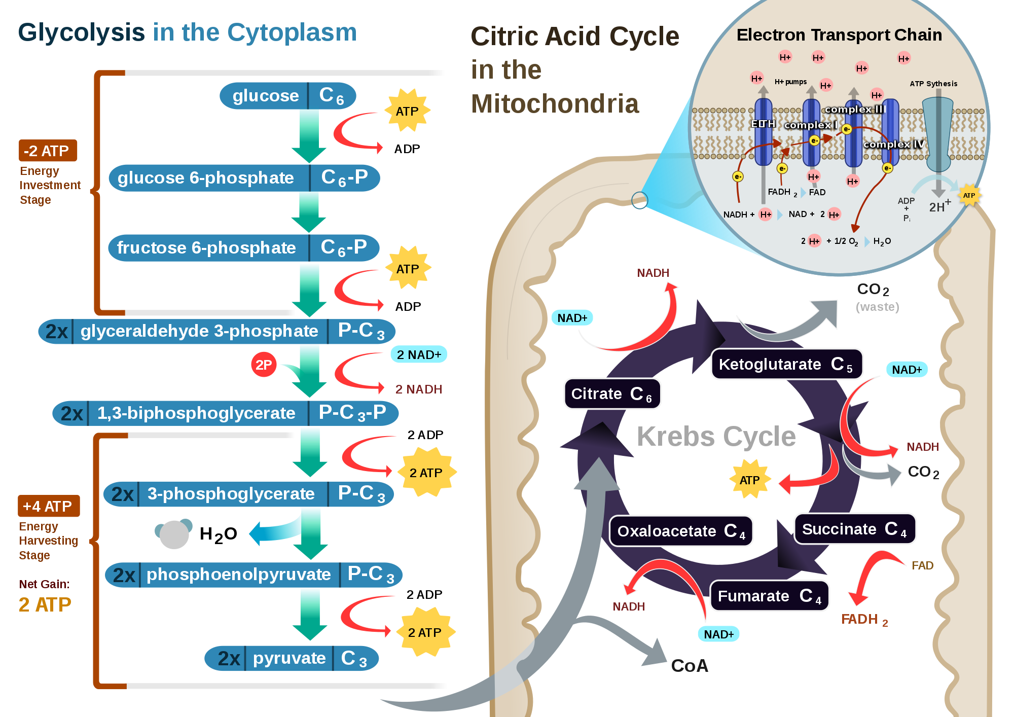
A much more detailed diagram of Cellular Respiration

Overall, this unit contained lots of information regarding the different organelles of eukaryotes, and focused a lot on photosynthesis and cellular respiration, the most complex topics of the unit. My only remaining questions are how prokaryotes function, since they have no organelles or nucleus.

Friday, October 16, 2015

Photosynthesis Virtual Labs

Photosynthesis Virtual Labs.

Lab 1: Glencoe Photosynthesis Lab


Analysis Questions
1. Make a hypothesis about which color in the visible spectrum causes the most plant growth and which color in the visible spectrum causes the least plant growth?
If chlorophyll is green, and reflects green light, then a plant exposed only to green light will not grow as well as a plant exposed to another color of light

If violet light has the shortest wavelength of all visible light, which has the most energy, then a plant exposed to violet light will grow better than a plant exposed to other colors of light.
2. How did you test your hypothesis? Which variables did you control in your experiment and which variable did you change in order to compare your growth results?
I grew both violet and green light exposed plants and compared them both to plants growing under red light as a control, and then compared the other colors of light to red.


Results:
Filter Color
Spinach Avg. Height (cm)
Raddish Avg. Height (cm)
Lettuce Avg. Height (cm)
Red


11 ⅔
Orange


6
Green


3 ⅓
Blue


12
Violet


8 ⅔

3. Analyze the results of your experiment. Did your data support your hypothesis? Explain. If you conducted tests with more than one type of seed, explain any differences or similarities you found among types of seeds.

My hypothesis was partially supported by my data. As I predicted, plants growing under green light grew the shortest of all the plants, but violet light didn’t cause the plants to grow the most. Instead, blue caused the most plant growth.


4. What conclusions can you draw about which color in the visible spectrum causes the most plant growth?
When grown under blue light, a plant will grow taller than if it was grown under a different color of light.

5. Given that white light contains all colors of the spectrum, what growth results would you expect under white light?

I would expect similar growth, because white light contains blue light, which caused the most plant growth.


Site 2: Photolab

This simulation allows you to manipulate many variables. You already observed how light colors will affect the growth of a plant, in this simulation you can directly measure the rate of photosynthesis by counting the number of bubbles of oxygen that are released.
There are 3 other potential variables you could test with this simulation: amount of carbon dioxide, light intensity, and temperature.
Choose one variable and design and experiment that would test how this factor affects the rate of photosynthesis. Remember, that when designing an experiment, you need to keep all variables constant except the one you are testing. Collect data and write a lab report of your findings that includes:
  • Question
  • Hypothesis
  • Experimental parameters (in other words, what is the dependent variable, independent variable, and control?)
  • Data table
  • Conclusion (Just 1st and 3rd paragraphs since there's no way to make errors in a virtual lab)
*Type this document on a word processor or in Google Docs and submit via Canvas.


In this lab we asked how temperature affected the rate of photosynthesis in plants. My hypothesis was that a plant subjected to higher temperatures would photosynthesize at a faster rate than a plant subjected to lower temperatures. To test this in the virtual lab, I set white light intensity and amount of Carbon Dioxide to the maximum allowed, and counted the amount of bubbles per minute at different temperatures, because without Carbon Dioxide and Light, photosynthesis would not occur.  My data showed that 25 degrees (C)  was the optimal temperature for photosynthesis because the most oxygen was produced at that temperature. This is probably because the enzymes in plants work best at that temperature, and anything lower or higher will begin to denature them.

This lab was done to demonstrate the effect on external conditions on internal functions such as photosynthesis. From this lab I learned how the rate of photosynthesis can be easily measured by counting the amount of oxygen bubbles, which helps me understand the reactants and products, such as oxygen, of photosynthesis and what happens to them. Based on my experience from this, I could choose optimal conditions for growing plants in a controlled environment.

Temperature Vs. Rate of Photosynthesis
Temperature (C)
Bubbles/Minute
10
24
25
66
40
54

Wednesday, October 7, 2015

Egg Diffusion Lab

Friday, October 2, 2015

Egg Macromolecules Lab Conclusion

In this lab, we asked which macromolecules, monosaccharides, polysaccharides, proteins, and lipids, if any, are found in the yolk, white, and membrane of an egg. We found that the egg yolk contained monosaccharides, the egg white contained monosaccharides, polysaccharides, proteins, and lipids, and the egg membrane contained monosaccharides, polysaccharides, and lipids. We tested for monosaccharides using benedicts solution, a solution that turns from blue to either green or orange in the presence of monosaccharides. When tested with the benedicts solution, the egg membrane turned dark blue, the egg yolk turned green-blue, and the egg white turned a different shade of blue, indicating monosaccharides in all 3. When testing for polysaccharides, iodine, which turns from brown to black in the presence of polysaccharides, was used to test for them. The iodine caused the egg membrane to turn dark brown, and the egg white to turn orange/light brown, which indicated polysaccharides in the egg membrane, and very few polysaccharides in the egg white. A mixture of Sodium Hydroxide (NaOH) and Copper Sulfate (CuSO) was used to test for proteins because when mixed with proteins, it turns from blue to purple. When mixed with all parts of the egg, only the egg white changed to a darker blue, which indicated proteins. Finally, we tested for lipids. For lipids, we used the chemical Sudan III, which changes from red to orange in the presence of lipids. Using that, the egg membrane and egg white turned orange, indicating lipids in both. The reason we found monosaccharides, polysaccharides, and lipids in the egg white was because they are used for energy by the developing organism. Proteins were found there because the organism can break them down and make its own proteins. The egg membrane had polysaccharides because they are used to communicate with other cells, and lipids were found because the membrane is made of phospholipids. Monosaccharides should not have been found, and were probably due to an experimental error. The yolk contained monosaccharides for energy.

Our data was unexpected due to various errors we made. When data from other identical experiments run in the class was compared, many people found macromolecules in parts of the egg that other groups didn't. In the egg membrane, we should have found proteins in addition to all of the other molecules we found because proteins are used during active transport in the egg membrane. In the egg yolk, we should have found polysaccharides for energy, lipids for the membrane around the yolk, and proteins for the developing organism. These errors were most likely caused by the color the egg yolk affecting the color of the chemical that was supposed to reveal the macromolecules. To fix this, less of the egg and more of the chemical should be used to test so the chemical reaction is more visible. Another error was in the lipid test. It was difficult to distinguish between the red that the chemical started as, and the orange it turned into because they are similar colors. To fix this, we could, again, use more of the chemical and less of the egg parts.

This lab was done to demonstrate where macromolecules are found in cells, and why there are there. From this lab I learned the purpose of different macromolecules, which helps me understand how cells carry out actions such as developing proteins and converting energy. Based on my experience from this lab, I can more accurately judge the effect of obvious errors in experiments, and have a better understanding on how cells work.